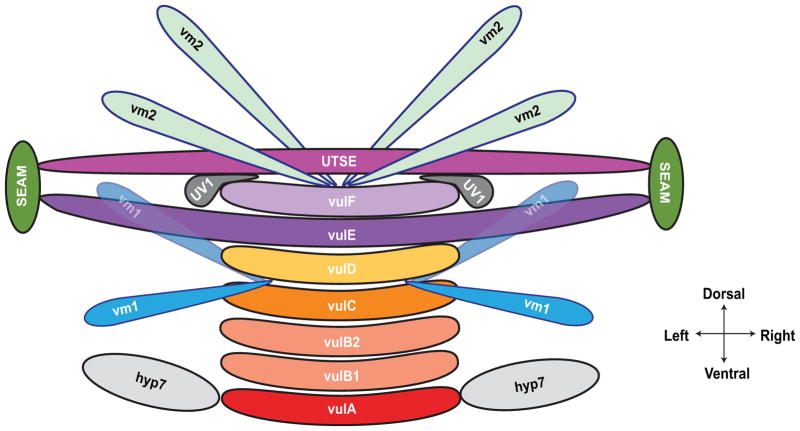
Figure 3.
Attachments of the vulva to other tissues. (A) The vulA cells on the ventral surface contact the hyp7 hypodermal syncytium; vulE cells extend laterally to contact the hypodermal seam cells; and vulF cells contact the uv1 cells and are overlaid by the utse (see Fig. 2F). Eight vulval muscles, four vm1 and four vm2, control vulval opening. vm1 cells contact the vulva between vulC and vulD toroids, and vm2 cells contact between vulF and the uterus. Modeled after 8, 102. For additional images of late vulval development, see WormAtlas 14.

