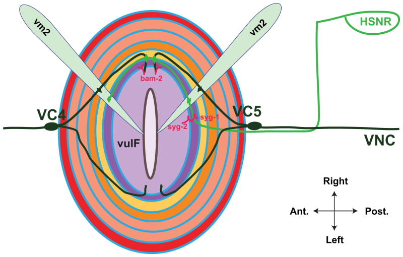
Figure 7.
Patterning of neuronal connections in the vulval region. Dorsal view of everted vulva. The two HSN neurons (shown is the right HSN in green) extend axons ventrally to the nerve cord, then anteriorly to the vulva. The interaction between HSN-expressed SYG-1 and vulF-expressed SYG-2 serves to guide the future sites of synaptic contact, which occur on vm2 muscle cells (shown are two of four vm2 cells, light blue) and VC neurons. VC4 and VC5 have cell bodies at the anterior and posterior of the vulval epithelium, respectively, and extend axons around the vulva and dorsally to synapse on vm2 cells. vulF-expressed BAM-2 is required to terminate axon branches. Modeled after 121 and 122.

