
Fig. 5.
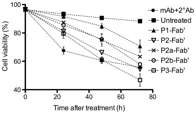
Time-dependent cell viability study assessed by propidium iodide (PI) binding. Treatment conditions were identical to apoptosis assays. Quantification was performed by flow cytometry. Experiments were carried out in triplicate (data shown as mean ± SD). (■) Untreated; (●) mAb+2°Ab; (▲) P1-Fab′; (▽) P2-Fab′; (×) P2a-Fab′; (○) P2b-Fab′; (□) P3-Fab′.