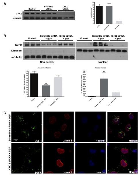
Fig. 4.
Knockdown of Clathrin blocks translocation of EGFR to the nucleus. (A) SKHep-1 cells were transfected with clathrin heavy chain 2 (CHC2) siRNA (50 nM). Immunoblots was performed to quantify the knockdown of CHC2 (left panel) and densitometric analysis confirms that it reduced protein expression by 85.4 ± 6.5% (n = 3, *p < 0.05) (right panel). Scramble siRNA was used and it didn’t reduce CHC2 expression level. (B) SKHep-1 cells were transfected with CHC2 or Scramble siRNAs and stimulated 48 h later with EGF (100 ng/mL) for 10 min. The amount of EGFR in the treated group with CHC2 siRNA and EGF didn’t alter in the non nuclear and in the nuclear fractions compared with control cells (p > 0.05). α-tubulin and Lamin B1 were used as purity controls for the non nuclear and nuclear fraction, respectively. Blot is representative of what was observed in nine separate experiments. (C) Confocal immunofluorescence images 10 min after stimulation with EGF (100 ng/mL). SKHep-1 cells were trasfected with Scramble or CHC2 siRNAs 48 h before the stimulation with EGF. EGFR and Lamin B1 were labeled with Alexa-488 (green) and Alexa-555 (red), respectively. Nuclear staining with Hoechst is shown in blue. Note that in CHC2 siRNA-treated cells the EGFR are concentrated outside the nucleus. Images are representative of what was observed in 20 cells under each experimental condition. **p < 0.001. Scale Bar = 10 μm.

