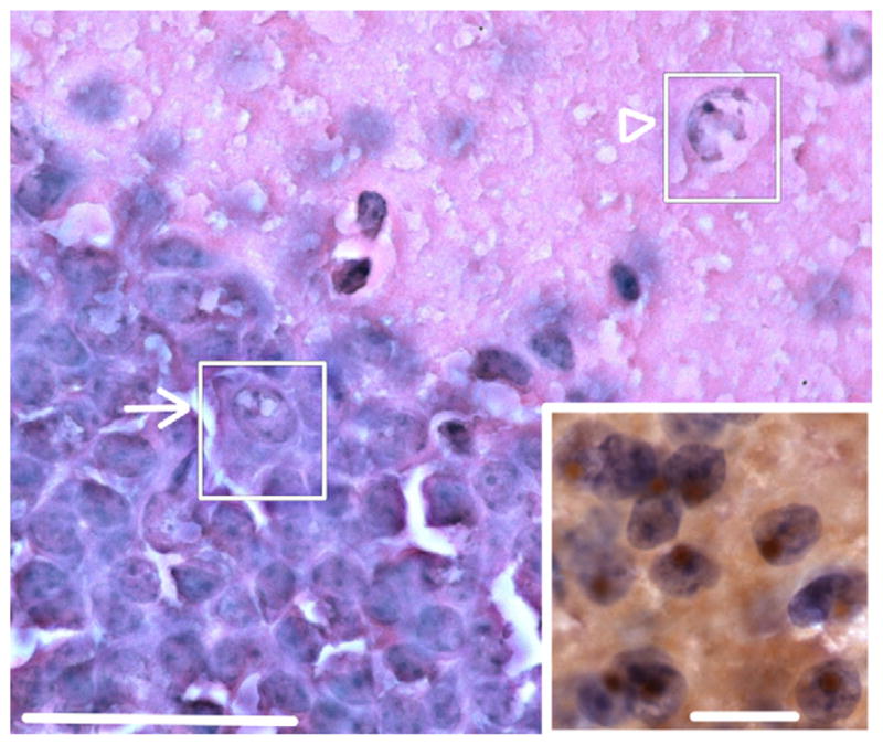
Fig. 2.

Intranuclear inclusions in dentate gyrus of female CGG KI mice. Hematoxylin and eosin (H&E) stained section of the dentate gyrus showing inclusions in granule cells (arrow; inset) and putative GABAergic cells in the inner molecular layer (arrowhead). Original magnification 1000 ×. Scale bar = 50 μm. Inset: numerous brown intranuclear inclusions by immunoperoxidase staining in hematoxylin stained cells of the posterior nucleus of the amygdala of a female CGG KI mouse. Original magnification 1000 ×. Scale bar = 10 μm. Main figure H&E stain, inset imunoperoxidase stain for ubiquitin. These images are from a female CGG KI mouse, 48 weeks of age with 12 repeats in the Fmr1 gene one X chromosome and 128 on the second Fmr1 gene. (For interpretation of the references to color in this figure legend, the reader is referred to the web version of this article.)
