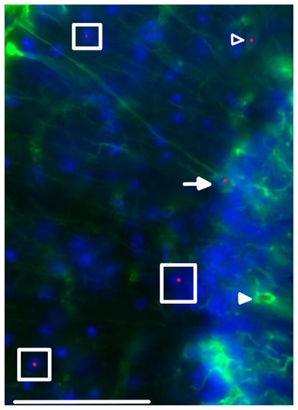
Fig. 4.

Intranuclear inclusion in astroglia and Bergmann glia. Protoplasmic (open arrowhead) and velate astroglial cells (filled arrowhead) stained for GFAP (green) and ubiquitin (red). Bergmann glial cell (arrow) stained for GFAP (green) and ubiquitin (red). Neurons negative for GFAP with intranuclear inclusions are shown with boxes. Cell nuclei stained with DAPI (blue). Arrowheads point to an astroglial cell in the inner molecular layer with an intranuclear inclusion as well as a velate astroglial cell in the granule cell layer with an intranuclear inclusion. Single plane confocal image. Original magnification 600 ×. Scale bar = 50 μm. This image came from a female CGG KI mouse, 55 weeks of age with 8 CGG repeats in the Fmr1 gene one X chromosome and 164 on the second Fmr1 gene. (For interpretation of the references to color in this figure legend, the reader is referred to the web version of this article.)
