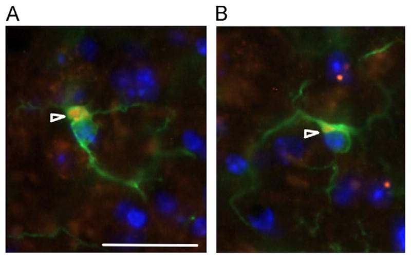
Fig. 5.

Intracellular masses in microglia. (A) Intracellular masses in CGG KI mice and (B) wildtype mice at 72 weeks of age. In microglia there appear to be intracellular, cytoplasmic masses (arrowheads) that stain positive for ubiquitin (red) as well as iba1 (green) in microglia. Nuclei stained blue with DAPI. These masses appear larger and less organized in wildtype mice (B) compared to female CGG KI mice (A). Original magnification 1000 ×. Scale bar = 10 μm. The image in A came from a female CGG KI mouse with 11 CGG repeats in the Fmr1 gene one X chromosome and 128 on the second Fmr1 gene and the image in B came from a wildtype littermate with both Fmr1 genes containing 10 CGG repeats. (For interpretation of the references to color in this figure legend, the reader is referred to the web version of this article.)
