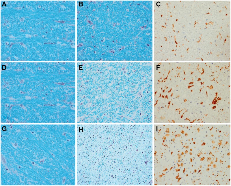
Figure 3.
Histological findings from a representative case from each of the three groups. A CSTD(−) case shows normal myelination of medial lemniscus and olivocerebellar fibres (A), normal myelination of corticospinal tract fibres (B), and only resting microglia, but no activated or phagocytic microglia (C). A CSTD(±) case shows normal myelination of medial lemniscus and olivocerebellar fibres (D), subtle evidence of myelin loss (E) with rare activated and phagocytic microglia (F). A CSTD(+) case shows normal myelination of medial lemniscus and olivocerebellar fibres (G), severe myelin loss (H) with numerous activated and phagocytic microglia (I). Luxol fast blue stain (A, B, D, E, G and H) and actin binding protein IBA-1 antibody (C, F and I). Sections taken at the level of the medullary pyramids. Magnification ×200 for all images.

