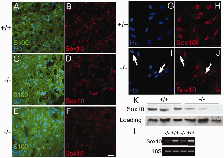
Figure 4.
Expression of SOX10 protein and messenger RNA is reduced in Merlin-null human schwannoma cells. (A–F) Cryostat sections of control human peroneal nerve (+/+, A and B) and human schwannoma tumours (−/−, C–F) stained with SOX10 antibody and the Schwann cell marker S100β (S100). Nuclei are counterstained with Hoechst dye (Ho). Almost all nuclei in control nerve (A and B) are SOX10 positive. For the tumour shown in panels C and D, a mosaic pattern is observed with many cells being SOX10 negative. For the tumour shown in panels E and F, almost no SOX10 stain is observed. Scale bar = 20 µm. (G–J) Immunolabelling of dissociated control Schwann (+/+, G and H) and Merlin-null schwannoma (−/−, I and J) cells in culture with SOX10 antibody. Note mosaic pattern in SOX10 stain in panel J; arrows show SOX10-negative cells in I and J. Scale bar = 20 µm. (K) Western blot analysis of three independent human control (+/+) and Merlin-null (−/−) schwannoma tumour samples. Note reduction of SOX10 expression in all tumour samples. (L) Semi-quantitative PCR showing reduction of SOX10 messenger RNA in two independent Merlin-null tumours (−/−) compared with normal Schwann cell controls (+/+).

