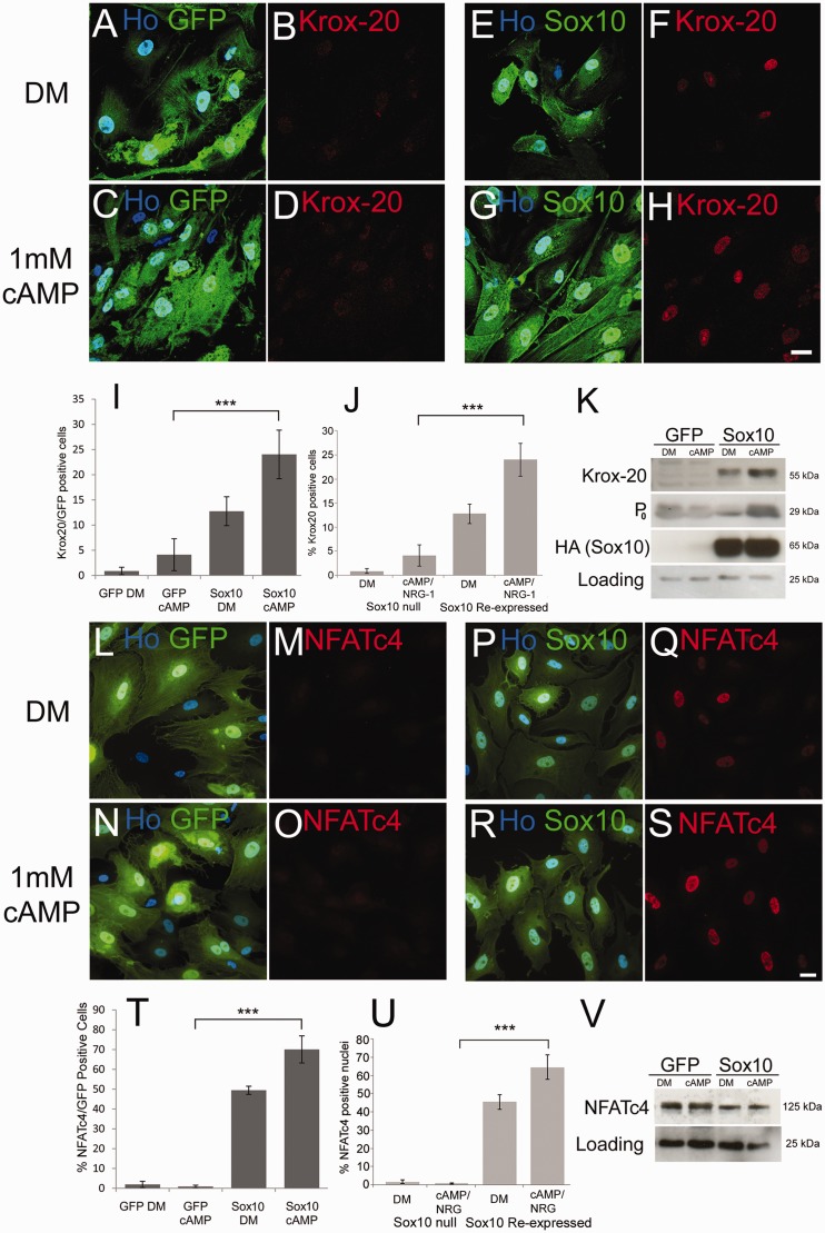
Figure 5.
Re-introduction of SOX10 into Merlin-null human schwannoma cells rescues induction of KROX20, P0 and NFATC4. (A–H) Merlin-null cells were infected with GFP (A–D) and GFP/SOX10 (E–H) expressing adenoviruses and either maintained in defined medium alone (A, B, E and F) or defined medium plus 1 mM cAMP (C, D, G and H). Note that SOX10 expression alone is sufficient to drive KROX20 expression in a small number of cells (F, I and K), which is further enhanced by cAMP treatment (H, I and K). Scale bar = 20 µm. (I) Percentage KROX20-positive cells in GFP- and SOX10-infected schwannoma cells. (J) Graph showing that SOX10-null mouse Schwann cells do not induce KROX20 in response to cAMP+NRG-1, but re-expression of SOX10 restores KROX20 induction. (K) Western blot showing regulation of KROX20 and P0 by SOX10 in Merlin-null cells. Probing with HA antibody detects adenovirally mediated SOX10 expression. (L–S) Re-expression of SOX10 in Merlin-null cells restores nuclear localization of NFATC4. Merlin-null cells were infected with GFP- (L–O) and GFP/SOX10- (P–S) expressing adenoviruses and either maintained in defined medium alone (L, M, P and Q) or defined medium plus 1 mM cAMP (N, O, R and S). Scale bar = 20 µm. (T) Percentage NFATC4-positive cells in GFP- and GFP/SOX10-infected schwannoma cells. (U) Graph showing that SOX10-null mouse Schwann cells do not induce nuclear localization of NFATC4 in response to cAMP+NRG-1, but re-expression of SOX10 restores NFATC4 relocalization. (V) Western blot showing expression of NFATC4 in GFP and GFP/SOX10 infected schwannoma cells. DM = defined medium.

