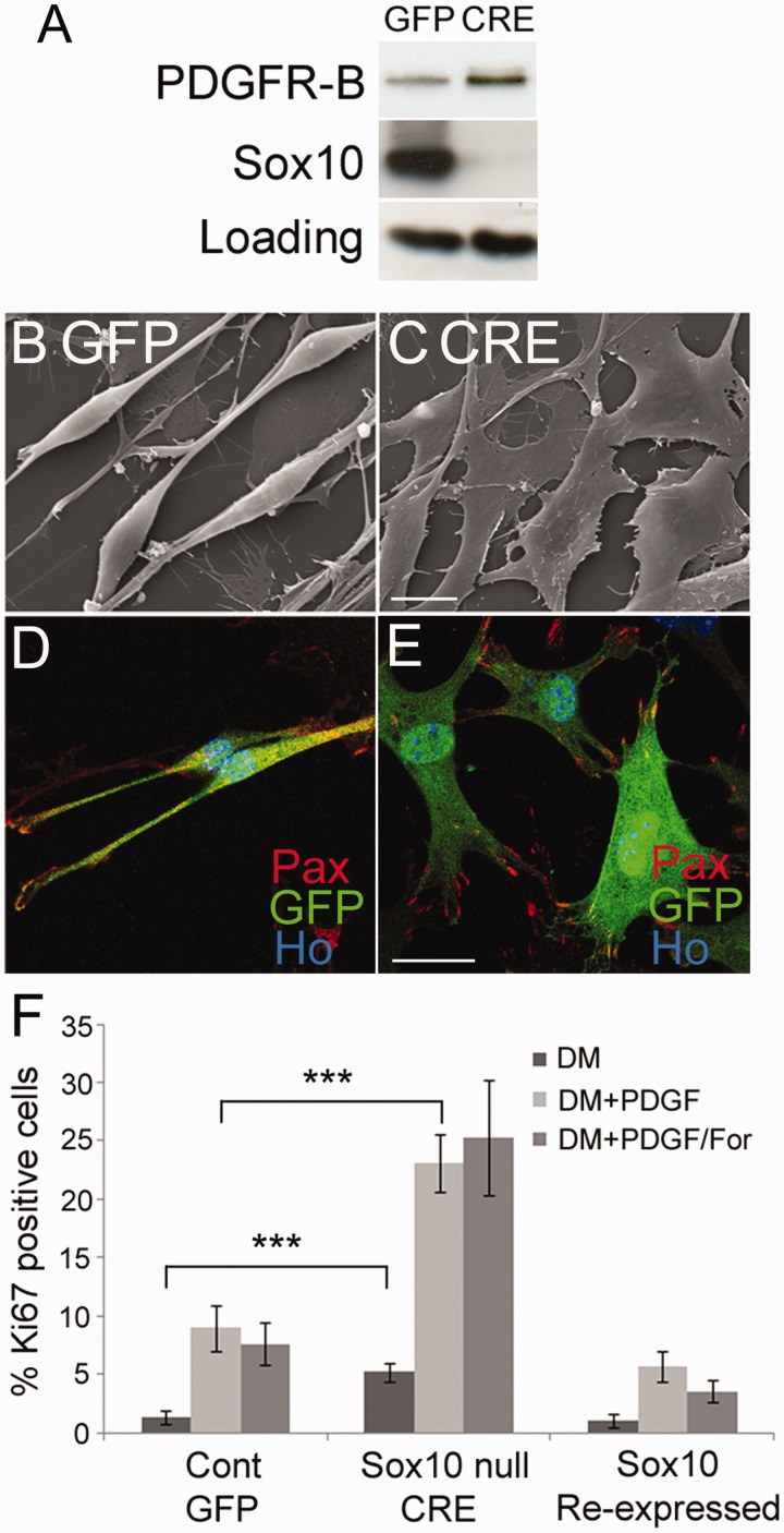
Figure 7.
Loss of SOX10 alone in mouse Schwann cells results in phenotypes typical of human schwannoma cells. (A) Western blot of SOX10fl/fl mouse Schwann cells infected with GFP- or Cre-expressing adenoviruses, showing increased expression of PDGFRB in SOX10-null mouse cells. (B and C) Scanning electron Micrsocopy showing flattening of SOX10-null cells (C) in comparison with control cells (B). Scale bar = 10 µm. (D and E) Immunolabelling of control (D) and SOX10 null (E) cells with paxillin antibody (Pax) to reveal increased numbers of focal adhesions in SOX10 null cells (E). Scale bar = 20 µm. (F) Graph showing significantly (P < 0.001) increased proliferation of SOX10-null mouse Schwann cells in response to 10 ng/ml of PDGF or PDGF plus 2 µM of forskolin (PDGF/For). Reintroduction of SOX10 into SOX10-null cells once more decreases PDGF-induced proliferation.

