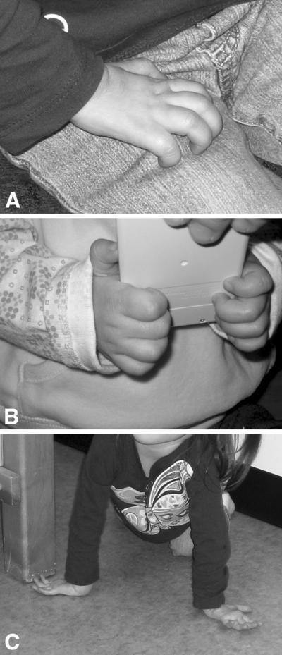
Figure 1.

Frozen sections of skeletal muscle biopsy from Twin A at 18 months of age stained with hematoxylin and eosin (A) as well as immunoperoxidase staining for slow (B) and fast (C) myosin heavy chain. A wide variation in fiber size (ranging from 5 – 40 μm in diameter) as well as a striking type I fiber predominance can be appreciated.
