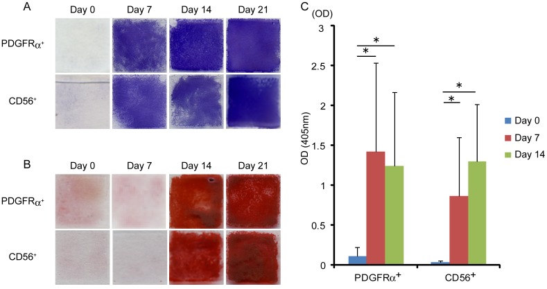
Figure 2. Osteogenic differentiation potential of human skeletal muscle-derived cells in vitro.
(A) Alkaline phosphatase staining was performed at the time points indicated during osteogenic differentiation of PDGFRα+ cells and CD56+ cells. (B) Alizarin red S staining was performed at the time points indicated during osteogenic differentiation of PDGFRα+ cells and CD56+ cells. (C) Alkaline phosphatase activity of PDGFRα+ cells and CD56+ cells during osteogenic differentiation was quantified. Values are shown as means ± s.d. of ten independent preparations. *P<0.01.

