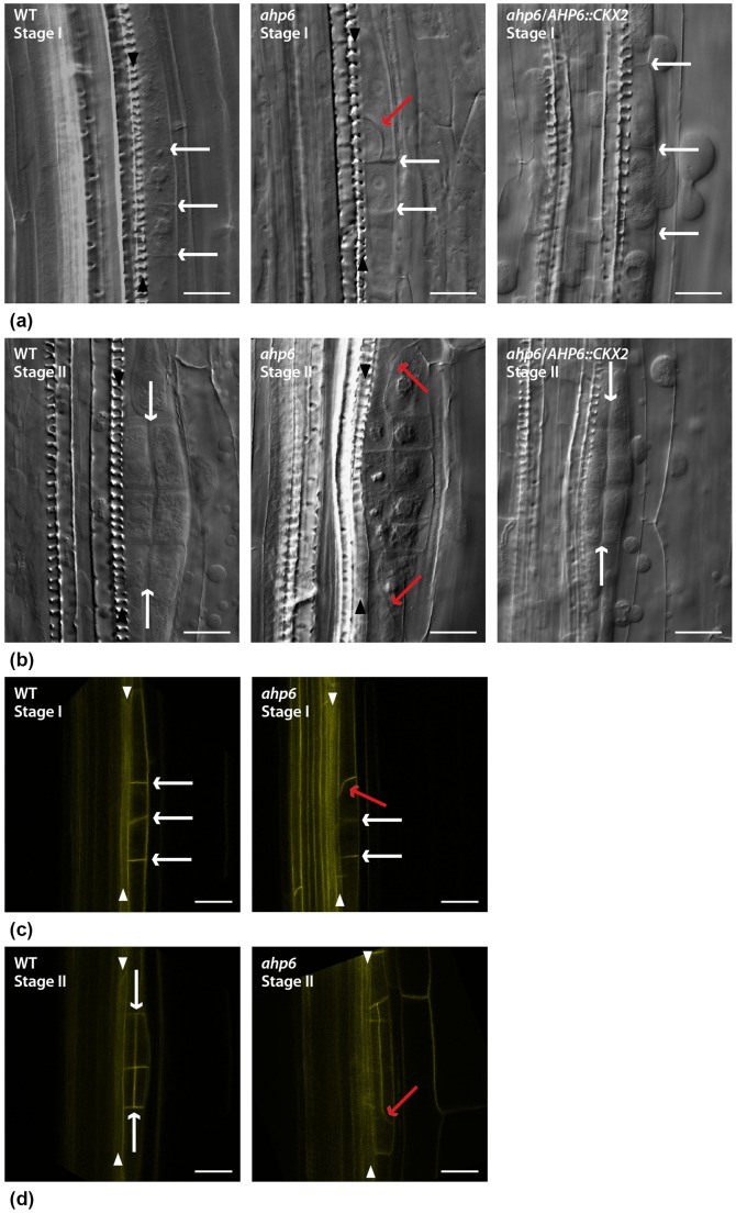
Figure 2. Abnormal cell division orientation of stage I and stage II ahp6 lateral root (LR) primordia.
a) Differential Interference Contrast (DIC) images of WT anticlinal pericycle founder cell divisions at stage I (white arrows) and a defective cell division (red arrow) in ahp6 at the same LR developmental stage; pericycle founder cell divided in the normal anticlinal orientation in ahp6/AHP6::CKX2 (white arrows). b) DIC images of WT and periclinal cell divisions of stage II (white arrows) and defective cell divisions (red arrows) in ahp6 at the same LR developmental stage; normal periclinal cell divisions in ahp6/AHP6::CKX2 (white arrows). c) AUX1-YFP as fluorescent marker to label the plasma membranes and show a WT stage I LR primordia anticlinal cell divisions (white arrows) and an abnormal cell division at stage I ahp6 primordia (red arrow). d) AUX1-YFP as fluorescent marker to label the plasma membranes and show a WT stage II LR primordia periclinal cell divisions (white arrows) and abnormal cell division orientation at stage II ahp6 primordia (red arrows). Arrowheads: Xylem cell files. Bars: 20 µm.

