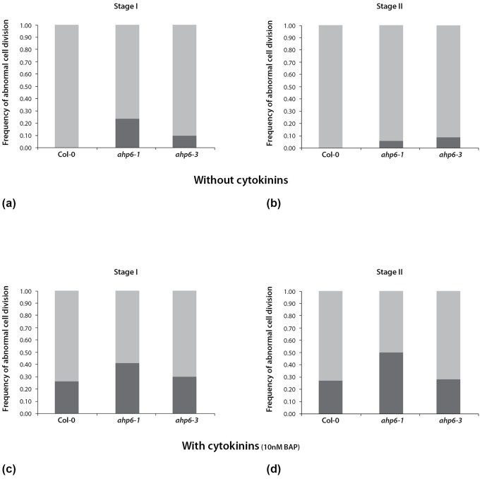
Figure 3. Frequency of abnormal cell divisions at stage I and stage II lateral root (LR) primordia.
a) At stage I, wild-type (WT) LR primordia (n = 40) show an invariant pattern where all cell divisions occur in an anticlinal orientation. In contrast, in LR primordia of ahp6 mutants at the same developmental stage show abnormalities in the plane of cell division: ≈25% for ahp6-1 (n = 37) and 10% for ahp6-3 (n = 48) LR primordia. b) At stage II, a similar invariant pattern of cell division was observed in the WT LR primordia (n = 61) with all cell divisions occurring in a periclinal orientation, whereas abnormally orientated cell divisions occurred in ≈5% for ahp6-1 (n = 72) and 10% for ahp6-3 (n = 55) LR primordia. This is data combined from three independent experiments with a total number of 58 WT roots, 84 ahp6-1 roots and 75 ahp6-3 roots. c) When grown with 10 nM cytokinin, about 25% of stage I WT LR primordia (n = 35) show abnormal cell divisions. There is also an additive increase in the number of abnormal cell divisions in CK treated ahp6-1 mutants with ≈40% of stage I LR primordia (n = 41) showing abnormal periclinal cell divisions. This effect is smaller in ahp6-3 where ≈30% of stage I LR primordia (n = 37) show aberrant cell divisions. d) When grown with 10 nM cytokinin, there is about 25% increase in the number of WT stage II LR primordia (n = 48) with abnormal orientation of cell divisions. The frequency of cell divisions with aberrant orientations is also increased in ahp6 stage II LR primordia: ≈50% for ahp6-1 (n = 46) and ≈30% for ahp6-3 (n = 53). This is data combined from two independent experiments with a total number of 51 WT roots, 58 ahp6-1 roots and 56 ahp6-3 roots.

