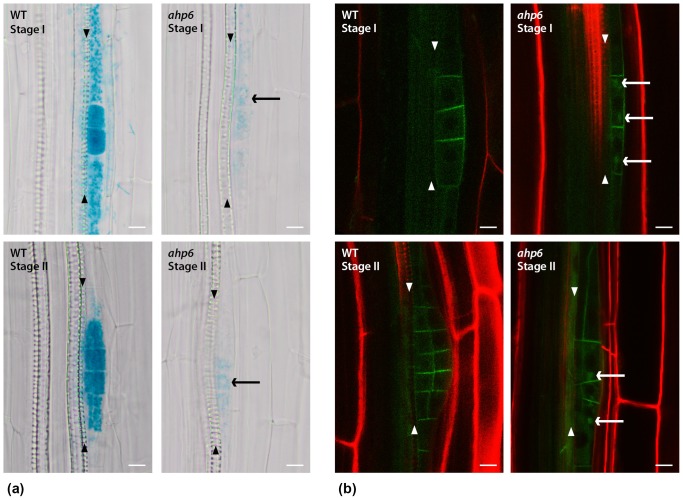
Figure 4. AHP6 and its interaction with auxin.
a) DR5::GUS signal is less intense in most of ahp6 lateral root primordia. Additionally the auxin response pattern is sometimes altered, for example: in some stage I and stage II LR primordia auxin response could only be observed in approximately half the cells (arrows point the stained half). b) PIN1-GFP is localized at plasma-membrane in LR primordia of WT and ahp6 mutant. Additionally, it shows an intracellular punctate pattern in around 35% ahp6 LR primordia (n = 56) (arrows). Arrowheads: Xylem cell files. Bars: 10 µm.

