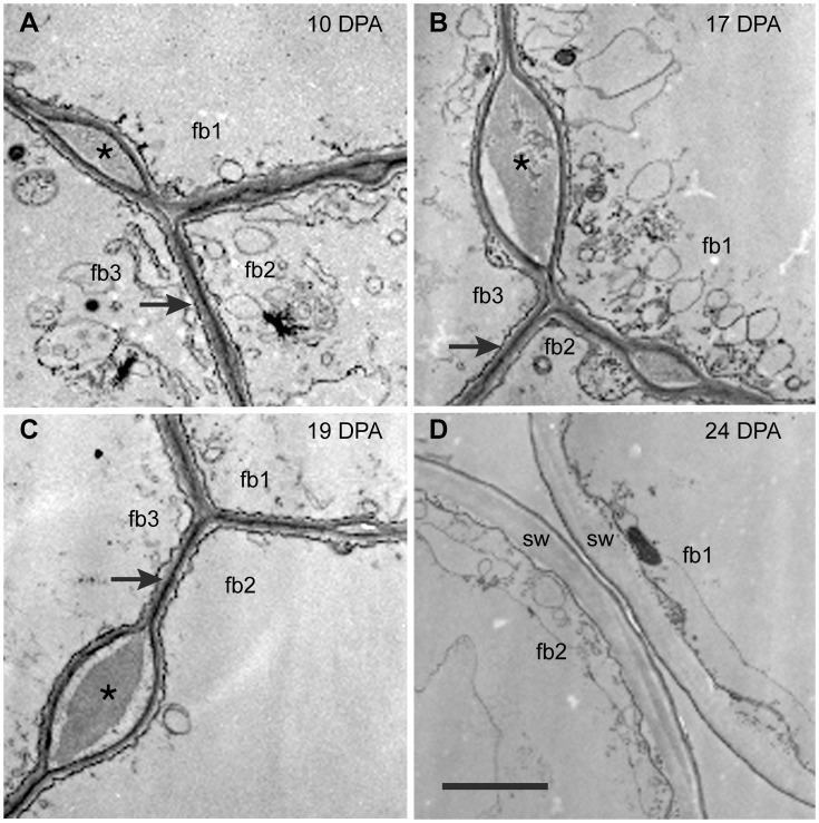
Figure 2. Transmission electron micrographs of cross-sectioned G. barbadense fiber at 10 to 24 DPA.
The ‘fb#’ labels indicate 2 or 3 individual fibers in each view. (A, B, C) At 10 DPA, 17 DPA, and 19 DPA, adjacent fibers are joined together by the CFML, the outermost layer of the primary wall. In many regions, a thin continuous wall exists between adjacent fibers (arrows in A, B, C). However, there are also periodic bulges between fibers that are filled with CFML material (asterisks in A, B, C). (D) At 24 DPA during secondary wall (sw) deposition, the fibers have separated due to CFML degradation, leaving empty space between them. The 2 µm scale bar in D applies to all micrographs.

