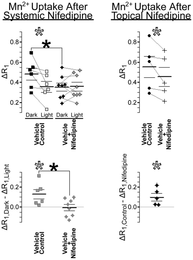
Figure 5. Effects of nifedipine on outer retinal Mn2+ uptake.

Scatter plots show outer retinal Mn2+ uptake (ΔR1; in s−1) in nifedipine-treated and vehicle control eyes. Inner retinal data are provided in Supplemental Figure S5 (in File S1). Paired measurements (left and right eye of each rat) are connected by grey lines in the top panels, and subtracted from one-another to produce the bottom panels. Left: In vehicle controls, dark-light differences are significantly greater than zero (open *s). Systemic treatment with nifedipine significantly inhibited Mn2+ uptake, but only in dark-adapted (patched) eyes (top filled *) – thereby significantly reducing dark-light differences (bottom filled *) to 0. Right: Mn2+ uptake in the dark-adapted outer retina was significantly lower in nifedipine-treated eyes than in the partner vehicle-control eyes (open *s).
