Figure 7.
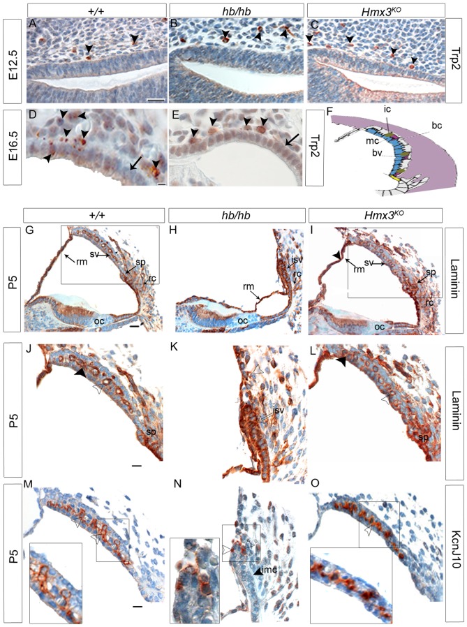
A–E: Trp2 expression performed on inner ear sagittal sections in hb/hb, Hmx3KO/Hmx3KO and control littermates at E12.5 and E16.5. A–C: Trp2 marks neural crest-derived melanoblasts at E12.5 (arrowheads in A). As shown by the arrowheads in B and C, we detect melanoblasts migrating from the neural crest to developing stria is detected in hb/hb, Hmx3KO/Hmx3KO, and littermate controls. D–E: While in control mice at E16.5 some intermediate cells precursors are already in the process of interdigitation in the developing stria vascularis (arrowheads in D), none are in the same process in the hb/hb mutants with only a couple of them lying outside the epithelium (arrowheads E). Moreover, Hematoxylin and Eosin counterstaining shows that the epithelium of developing stria looks immature in hb/hb compared to littermate controls (arrows in D,E). Scale bars: 10 µm. F: Cartoon of the cellular structure of stria vascularis. Adapted from [77]. G–I: Immunohistochemistry showing Laminin expression in cochlea and general cochlear morphology of hb/hb, Hmx3KO/Hmx3KO and control littermates at P5. Laminin is expressed in all cochlear basal lamina [63], including stria vascularis, Reissner's membrane, root cell processes and spiral prominence (arrows in G). J–L: Laminin expression in stria vascularis of hb/hb, Hmx3KO/Hmx3KO and control littermates at P5. At this stage, we detect basal lamina in blood vessel endothelia (arrowhead in J) and in very small pockets below marginal cells (black arrowhead in J) in control mice. In hb/hb we detect a much stronger laminin expression (denser basal lamina) around the immature stria vascularis (arrow in K). Moreover, we observe fewer and smaller blood vessels in hb/hb compared to littermate controls (examples of blood vessels are labeled with transparent arrowheads in J,K,L). We did not detect any difference in laminin expression in Hmx3KO/Hmx3KO compared to littermate controls (arrowhead in L). M–O: Kcnj10 expression in the stria vascularis of hb/hb, Hmx3KO mutants and control littermates at P5. Kcnj10 is an inward potassium channel of intermediate cells. We detect only some intermediate cells in hb/hb mutants (arrowheads in N) compared to their littermate controls at this stage (M). These intermediate cells are just outside the undifferentiated strial epithelium (the black arrowhead in N points to the immature marginal cell layer in hb/hb, see also Figure S1). No difference in Kcnj10 expression is detected in Hmx3KO/Hmx3KO at this stage compared to control littermates (arrowhead in O), consistent with their EP values being close to normal. Boxes delimit regions in higher magnification. Scale bars: A–E: 20 µm; G,I: 10 µm; J–O; 20 µm. bc: basal cells, bv: blood vessels, ic: intermediate cells, imc: immature marginal cells, isv: immature stria vascularis, oc: organ of Corti, rc: root cell processes, Rm: Reissner's membrane, sp: spiral prominence, sv: stria vascularis.

