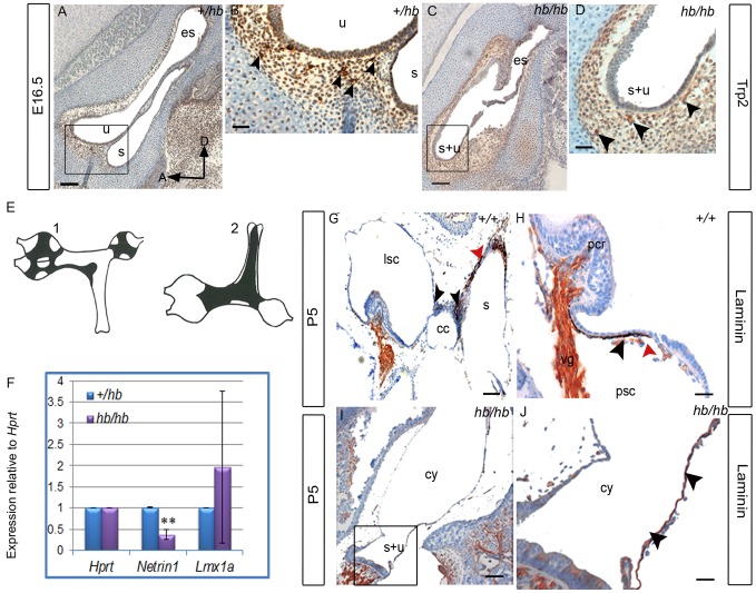
Figure 9. The headbobber stria vascularis remains immature with lack of intermediate cells and impaired basal lamina degradation.
A–D: Trp2 expression in the vestibular system at E16.5 is analysed by immunohistochemistry in the hb/hb and control littermates on sagittal sections. At this stage, melanoblasts have already reached all the different vestibular structures (arrowheads in B, see E). We can still detect amelanoblasts in hb/hb vestibular system (arrowheads in D). Scale bars: A,C: 100 µm; B,D: 20 µm. Boxes delimit regions in higher magnification. E: Schematic drawings of the flattened vestibular membranes of normally pigmented mice showing distribution of melanocytes in the vestibule viewed on medial (E1) and lateral (E2) position, adapted from [7]. F: Quantitative real-time PCR on cDNA generated from RNA from E12.5 littermate embryo half heads. Netrin-1 mRNA levels are significantly lower in hb/hb than in littermate controls. Error bars, s.d. Quantity normalised to Hprt1 levels. N = 3. **: p<0.01. G–J: Laminin staining in the vestibular system of hb/hb and littermate controls at P5. In controls, we find melanocytes between the common crus and the other vestibular structures (black arrowheads in G) as well as on the saccular wall (red arrowhead in G) and adjacently to cristae (black arrowhead in H). We show that basal lamina is breaking close to melanocytes (red arrowhead in H). We cannot detect any distinct structure nor melanocytes in the vestibular system of hb/hb mutants at this stage (I,J), but we detect the presence of an immature and tight basal lamina all around the vestibular cyst wall, where melanocytes are supposed to be positioned. (black arrowheads in J compared to H). Scale bars: 10 µm. A: anterior, cc: common crus, cy: vestibular cyst, D: dorsal, es: endolymphatic sac, lsc: lateral semicircular canal, pcr: posterior cristae, psc: posterior semicircular canal,,s: saccule, s+u: utriculosaccular space, u: utricle.

