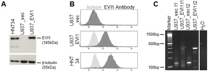
Figure 1. Establishment and characterization of a human myeloid cell line constitutively overexpressing EVI1.

A) Immunoblot analysis for the detection of EVI1 in U937_vec and U937_EVI1 cells. HNT-34 cells, which express EVI1 due to a rearrangement of chromosome band 3q26 [43], were included for comparison. Hybridization with a β-tubulin antibody was used as a loading control. B) Intracellular FACS staining for detection of EVI1 in U937_vec, U937_EVI1, and HNT-34 cells. Dark grey histogram curves, EVI1 antibody; light grey histogram curves, isotype control. C) High-resolution gel analysis of LAM-PCR amplicons obtained from Tsp509I digested genomic DNA from U937_vec and U937_EVI1 cells. DNA was isolated shortly after infection and sorting (t1) as well as after another 12–15 weeks in culture (t2).
