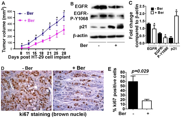
Figure 5. Berberine exhibits an anti-tumor effect on the xenograft model.
Nude mice bearing HT-29 xenografts were treated with berberine (0.1% in drinking water) starting at the day of inoculation until the end of the experiment. Tumor volumes were measured at the indicated days post HT-29 cell implant (A). Tumor tissues were prepared for Western blot analysis of indicated signaling pathways. Anti-β-actin antibody was used as a protein loading control (B). The fold change of the band density was determined by comparing the density of indicated band to the β-actin band of the same mouse (C). Tumor tissues were fixed and prepared for immunohistochemistry of ki67. Brown nuclei represent ki67 positive staining (D). The percentage of ki67 positive staining cells was quantitatively analyzed using Ariol SL-50 automated slide scanner (E). In C, * p<0.05 compared the control mice. In D, * p<0.01 compared the corresponding group in control mice. N = 7 mice in each group.

