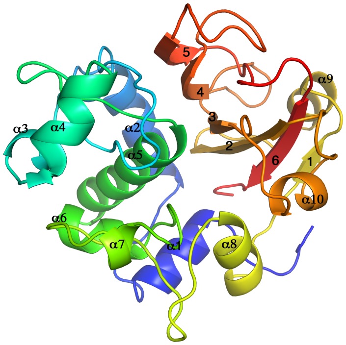
Figure 3. Cartoon diagram of the overall three-dimensional structure of NlpC/P60_2.
Rainbow-colored α-helixes and β-strands are shown from blue at the N-terminus to red at the C-terminus. The β-sheet (formed by antiparallel β-strands 1-6-2-3-4-5) is flanked by short α-helixes (3-4-7-9) and a globular α-helical domain (formed by α-helixes 1-2-5-6). All 10 α-helixes are located at the N terminus, followed by the six β-strands.

