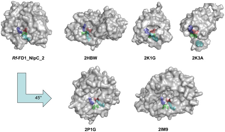
Figure 8. Location of the active-site grooves of various members of the NlpC/P60 superfamily represented on the molecular surface (annotated by PDB entry number).
The catalytic triad residues are colored as follows: cysteine in red, histidine in blue, and the third polar residue in green. The active-site grooves of 2HBW, 2K1G, 2K3A are located on the same face as that of NlpC/P60_2. The active-site residues of all structures were first superimposed; in order to view the grooves of 2P1G and 2IM9. The latter structures were then rotated approximately 45° counter-clockwise, relative to NlpC/P60_2.

