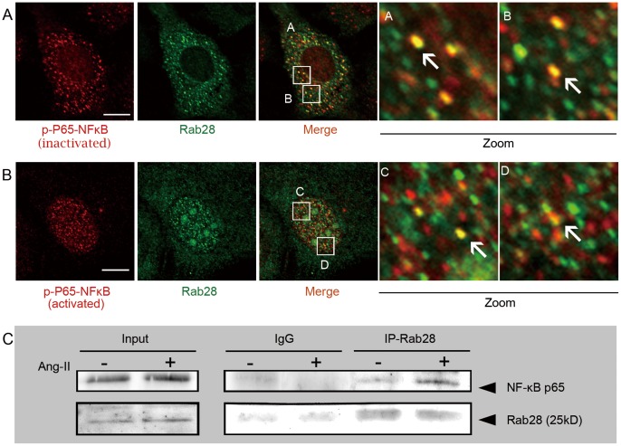
Figure 5. Co-localization and co-immunoprecipitation of NF-κB and Rab28 in ECs.
(A) NF-κB and Rab28 were co-localized, at least partly, in the cytoplasm of starved ECs. (B) Stimulation of ECs with exogenous angiotensin II (Ang II) caused the translocation of both Rab28 and activated NF-κB into the nucleus, with these two molecules still partially co-localized. Scale bars: 10 µm. (C) Whole cell lyastes of ECs treated with 10−6 M Ang II were prepared and probed for co-immunoprecipitation assays with anti-Rab28 antibody. Proteins from the immunoprecipitateds were detected by Western blot using anti-NF-κB p65 antibody. NF-κB co-immunoprecipitated with Rab28.

