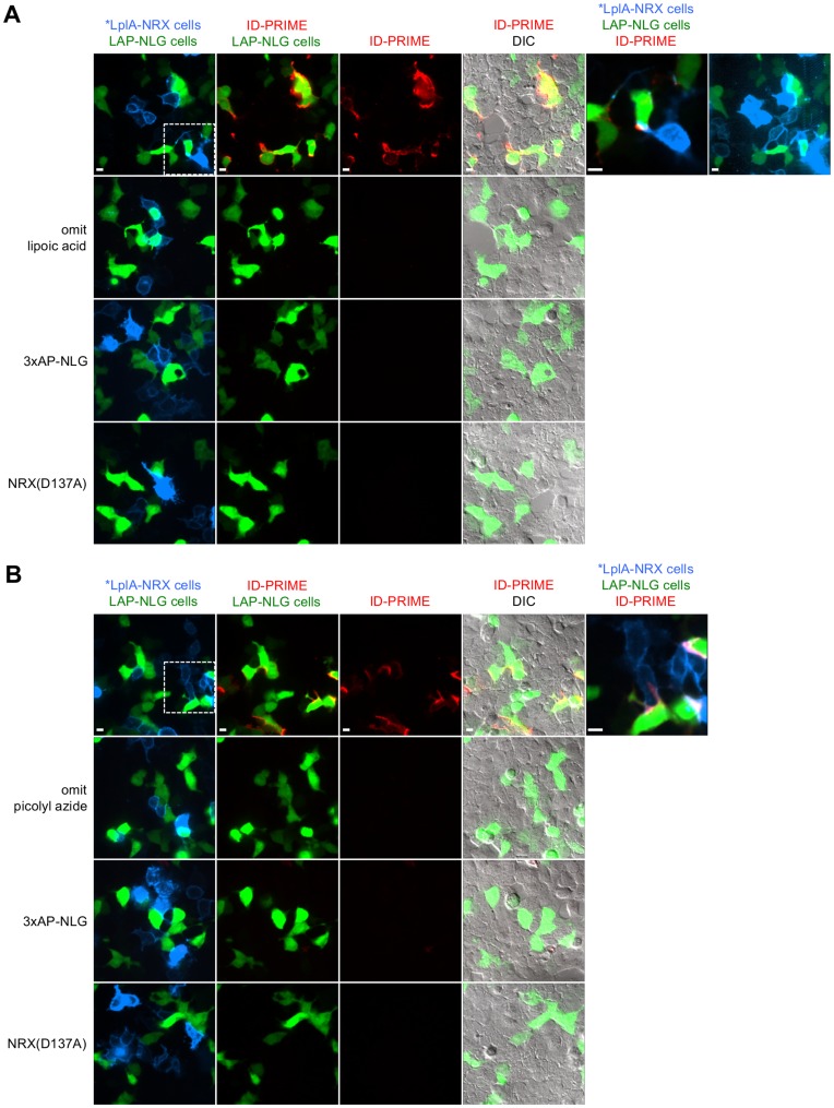
Figure 4. ID-PRIME for imaging neurexin-neuroligin interactions in HEK cells.
(A) ID-PRIME with lipoic acid readout (as in Figure 1B). HEK cells were separately transfected with *LplA36-NRX3β plus a membrane-localized tdTomato marker (shown in blue), or 3xLAP-NLG1 plus a Venus marker. After mixing and replating, cells were labeled with 50 µM lipoic acid +500 µM ATP for 15 minutes. Ligated lipoic acid was detected with an anti-lipoic acid antibody followed by a secondary antibody-AF647 conjugate (shown in red) for 5 minutes each. For row 1, a magnified view representing the boxed region, and a more contrasted view of the transfection markers are shown on the right. Controls were performed with lipoic acid omitted (row 2), the acceptor peptide for BirA substituted for LAP (row 3), and the interaction-deficient NRX mutant (row 4). (B) ID-PRIME with picolyl azide readout (as in Figure 1C). HEK cells were transfected as in (A), and labeling was performed with 100 µM picolyl azide +500 µM ATP for 15 minutes, followed by detection with copper-catalyzed click chemistry, using 50 µM copper and 20 µM alkyne-AF647. Color schemes and controls are the same as for (A). All scale bars, 10 µm.

