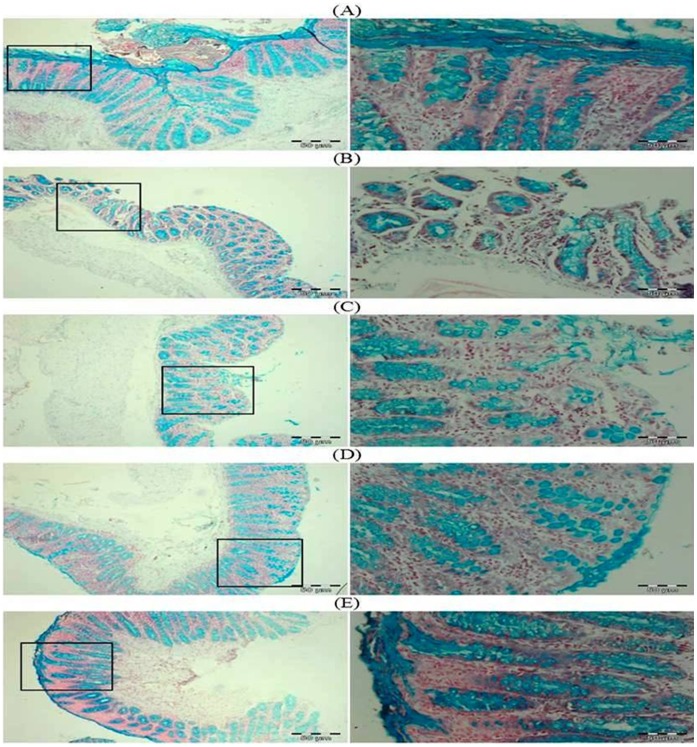
Figure 6. Photomicrographs depicting mucin staining.
In DMH treated group (Group II), there is regional depletion of mucin in the form of mucous layer (blue in color). Treatment with glycyrrhizic acid decreased the depletion of the mucous layer in Group IV as compared to Group II. No effects of glycyrrhizic acid on mucous layer in Group III as compared to Group II. There is no depletion of the mucous layer in colonic sections of Group I and Group V. Insets at the right panel show a magnified view (40x magnifications) of the insets showed at the left panel (10x magnifications).

