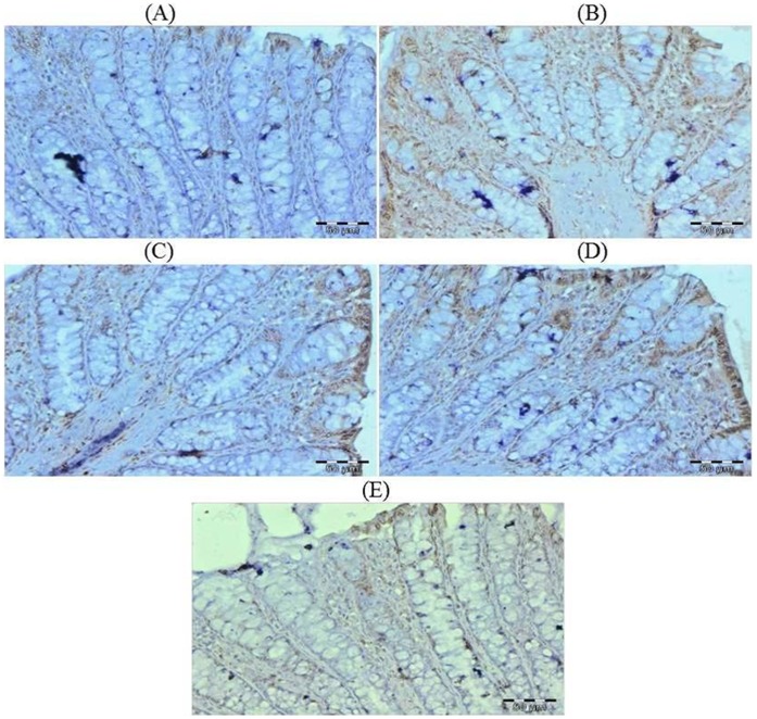
Figure 9. Photomicrographs depicting immunohistochemical staining of Ki-67.
For immunohistochemical analyses, brown colour indicates specific immunostaining of Ki-67 and light blue colour indicates nuclear haematoxylin staining. The colonic section of DMH-treated group (Group II) has more Ki-67 immunopositive staining as indicated by brown colour as compared to control group (Group I) while treatment with glycyrrhizic acid in Group III and Group IV reduced Ki-67 immunostaining as compared to Group II. However there was no significant difference in the Ki-67 immunostaining in Group V as compared to Group I. Original magnification: 40x.

