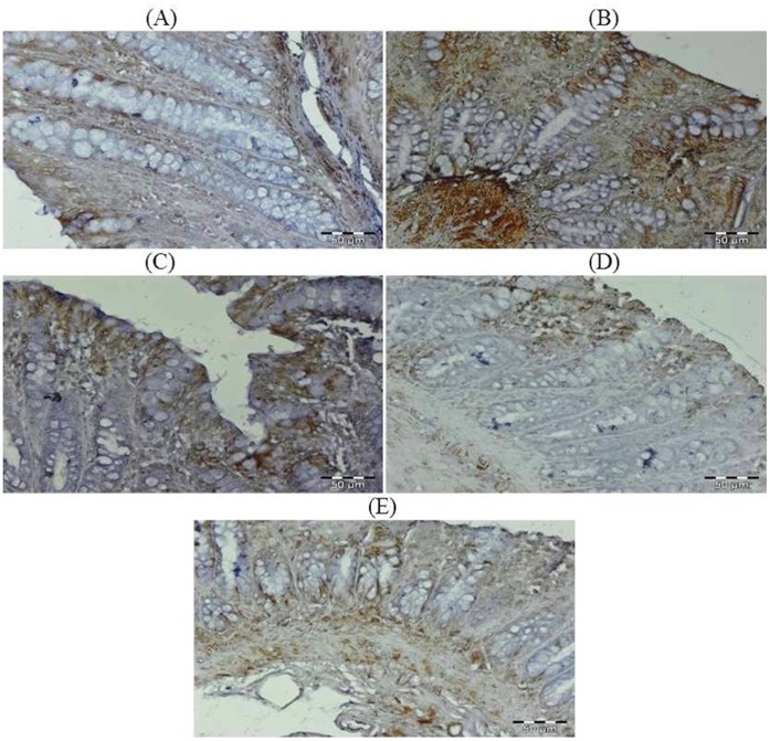
Figure 13. Photomicrographs depicting immunohistochemical staining of VEGF.
For immunohistochemical analyses, brown colour indicates specific immunostaining of VEGF and light blue colour indicates nuclear haematoxylin staining. The colonic section of DMH-treated group (Group II) has more VEGF immunopositive staining as indicated by brown colour as compared to control group (Group I) while treatment with glycyrrhizic acid in Group III and Group IV reduced VEGF immunostaining as compared to Group II. However there was no significant difference in the VEGF immunostaining in Group V as compared to Group I. Original magnification: 40x.

