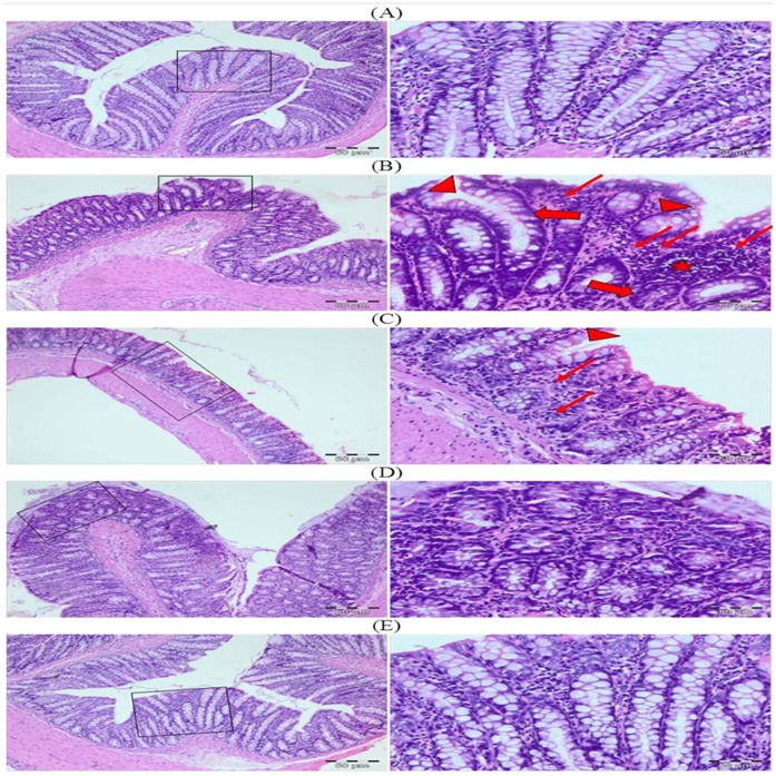
Figure 19. Photomicrographs depicting histology of rat colon.
The histological sections of control group showed normal histoarchitecture while DMH treated group (Group II) exhibited intense inflammatory cells infiltration, irregular glandular structure along with crypt ablation. In Group III and Group IV, histological sections showed that treatment with glycyrrhizic acid showed protection against DMH induced colonic damage. Colonic sections of Group V (only glycyrrhizic acid treated group) displayed normal histology as similar to that of Group I (control group). Insets at the right panel show a magnified view (40x magnifications) of the insets showed at the left panel (10× magnifications).

