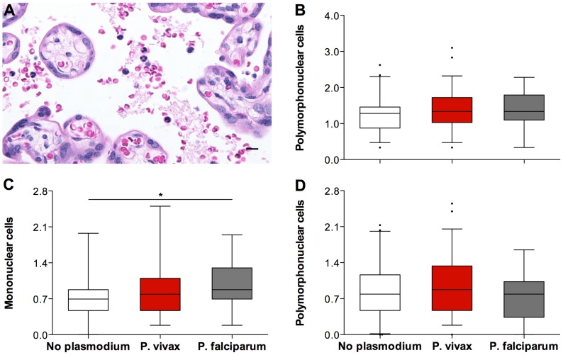
Figure 3. The immune-cell parameters evaluated by Plasmodium species during infection.
The percentage of immune cells present in the intervillous space of the placentas evaluated (A) was calculated after counting a total of 500 intervillous space cells. Total leucocytes percentage (B), mononuclear cells percentage (C) and polymorphonuclear cells percentage (D) were plotted against Plasmodium exposure during pregnancy, assessed by microscopy. The placentas from the “no plasmodium” group (n = 41; white boxes) appear to have less immune cells present in the intervillous space than the placentas from the “P. vivax” group (n = 59; red boxes) and the placentas from the “P. falciparum” group (n = 19; grey boxes). * ANOVA test, P-value = 0,039. Graphs (B, C, and D) represent the transformed data. The boxes represent the mean and standard deviation values. The whiskers represent the 5th and 95th percentiles. The photograph was taken using a Zeiss Axio Imager M2 light microscope equipped with a Zeiss Axio Cam HRc. Grid overlays and counts were performed using Image J.

