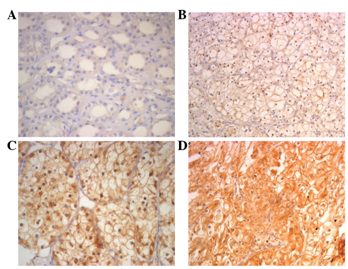
Figure 2.

Immunohistochemical analysis of the expression of FER protein. FER is mainly localized within the nuclei and cytoplasm. (A) Immunostaining of the ADT samples was negative or at a very low level. (B) Weak FER staining in cancerous tissue. (C) Moderate FER staining in cancerous tissue. (D) Strong FER staining in most of the tumor cells. Magnification, ×400. ADT, normal adjacent tissues.
