
Figure 1.
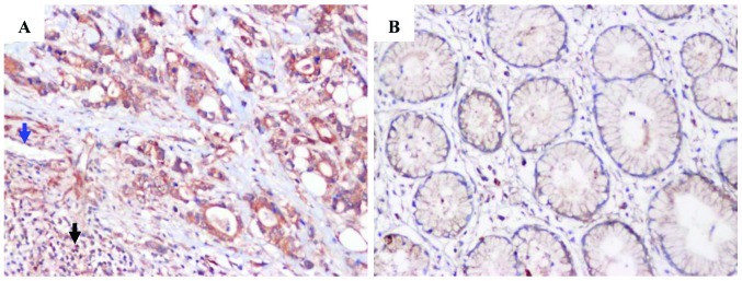
Representative immunohistochemical staining for CXCR1/2 in corresponding non-neoplastic mucosa tissue and tumor. (A) Tumor tissue with strong expression. CXCR1/2 were also present in some leukocytes (black arrowhead) and vascular endothelial cells (blue arrowhead). (B) Corresponding non-neoplastic mucosa tissue. Original magnification, x200. IgG stainingwas used as a negative control. CXCR1/2, C-X-C chemokine receptor types 1/2.