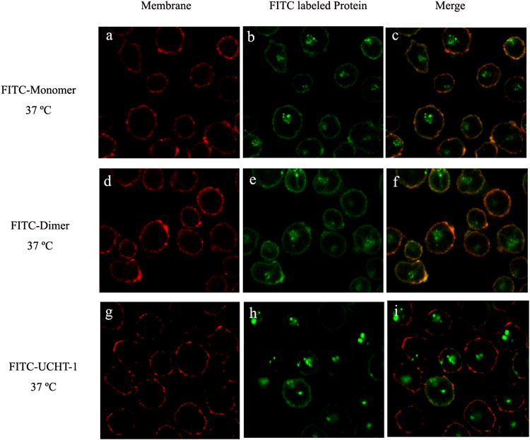
Figure 2.

Confocal images of intracellular distribution of FITC-labeled anti-CD3-DHFR2 monomer, dimer or UCHT-1 at 37°C. HPB-MLT cells were incubated with FITC-labeled anti-CD3-DHFR2 monomer (a-c), FITC-labeled anti-CD3-DHFR2 dimer (d-f), or FITC-labeled UCHT-1 (g-i) for 2 hours at 37°C. Cell membranes were stained with concanavalin A-Alexa Fluor 594 (Red). Superposition of red channel and green channel were shown in images c, f, and i.
