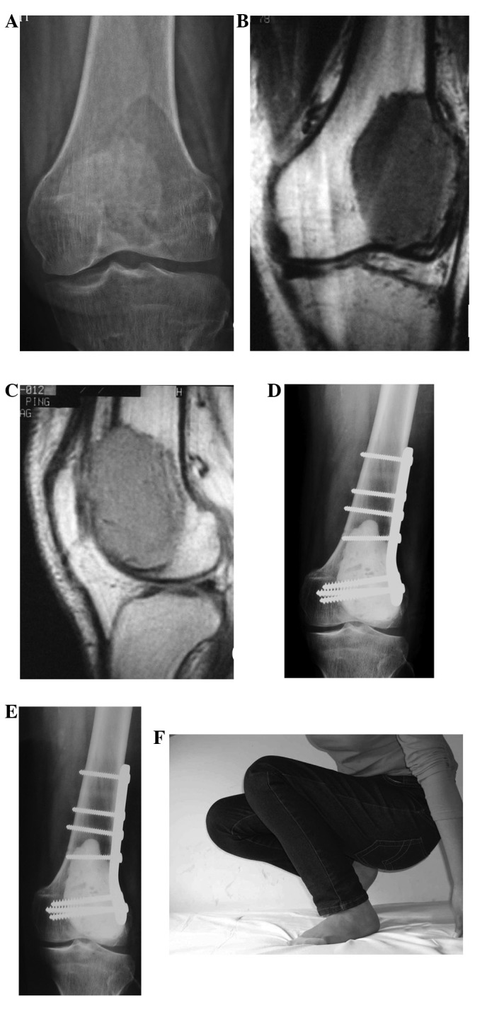
Figure 2.

A 30-year-old female patient with giant cell tumor (GCT) of the left distal femur. (A) X-ray film reveals osteolytic destruction and cortical thinning at the left femoral condyle. (B) and (C) T1-weighted MRI reveals low signal at the distal femur and a lateral visible tumor penetrating the front side of the cortex. (D) 4 days after surgery, postoperative X-ray reveals bone cement filling and internal fixation are normal. (E) and (F) 22 months after surgery, X-ray shows that the joint space is normal, no lucent zones surround the bone cement, internal fixations are firm and the joint function is normal.
