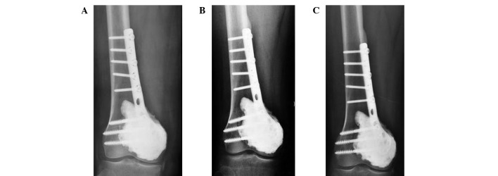
Figure 3.

A 41-year-old male patient with GCT of the right distal femur. (A) 4 days after surgery, X-ray film reveals bone cement filling and internal fixation are good. (B) 12 months after surgery, X-ray film shows the bone cement surrounding the lucent zones, normal joint space and internal fixation. (C) 31 months after surgery, the bone cement surrounds lucent zone with no progression.
