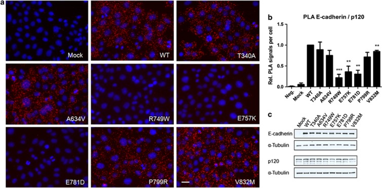
Figure 2.
Changes in the p120 binding domain are dramatic for E-cadherin and p120-catenin association. (a) The interaction between WT or mutant E-cadherin and p120-catenin was assessed by in situ PLA. CHO cells stably transfected with the empty vector (Mock) or with WT, T340A, A634V, R749W, E757K, E781D, P799R or V832M hE-cadherin were fixed and incubated with antibodies against E-cadherin and p120-catenin. In the negative control (Neg.), CHO WT cells were incubated only with the antibody against E-cadherin. Close proximity of oligonucleotide-ligated secondary antibodies allows rolling-circle amplification and detection of the amplification product by a fluorescence labeled probe (red dots). Nuclei were counterstained with DAPI (blue). The pictures were taken under a × 40 objective. Scale bar represents 20 μm. (b) The number of PLA signals per cell was quantified in each condition. The graph shows the average of relative number of PLA signals per cell±SE, n=3 (**P≤0.01 and ***P≤0.001). (c) E-cadherin, p120-catenin and α-Tubulin expression levels were detected in whole-cell lysates by western blot. α-Tubulin was used as a loading control. The images shown are representative of three independent experiments.

