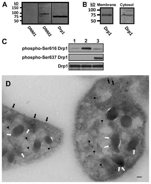
Figure 1. Detection and localization of Drp1 in platelets.
(A) Human platelet lysates were evaluated for dynamin 1 (DNM1), dynamin 2 (DNM2), and dynamin-related protein-1 (Drp1) as indicated. (B) Platelet cytosol and membraneswere evaluated for Drp1. (C) Platelet were incubated with either 1) buffer alone, 2) 5 μM SFLLRN, or 3) 10 μM forskolin. Lysates were then evaluated using either an anti-Ser616 Drp1 phosphorylation site specific antibody (phosphor-Ser616 Drp1), an anti-Ser637 Drp1 phosphorylation site specific antibody (phosphor-Ser637 Drp1), or an antibody to detect total Drp1 (Drp1). (D) Immunogold staining of resting platelets demonstrates Drp1 associated with plasma membrane (black arrows), granule membranes (white arrows), and cytosol (black arrowheads). Scale bar, 100 nm.

