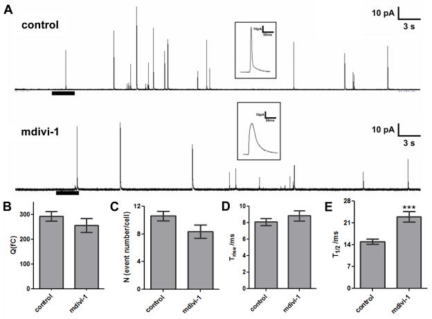
Figure 4. Role of Drp1 in the kinetics of platelet granule release.
(A) Representative amperometric traces of rabbit platelet dense granule release in the presence and absence of mdivi-1. The heavy bar beneath the tracings indicates the time and the duration of the thrombin stimulation. Insets: The distinct spike shapes for control (upper) and mdivi-1 (lower) conditions depicted in the millisecond time scale demonstrate different serotonin release kinetics. Mdivi-1 (10 μM) did not influence quantal release (B), the number of granules released per cell (C), or the time required for transition from fusion pore to full fusion (Trise) (D). However, the total time required for the release event (T1/2) was longer for platelets exposed to mdivi-1 (p<0.001) (E). Data represent the average ± S.E.M. of 107 control tracings and 39 tracings from platelets exposed to mdivi-1.

