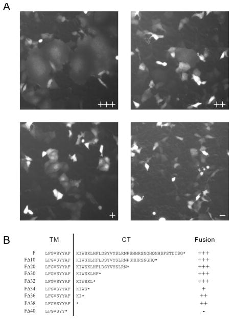
Figure 1.
Functional characterization of TPMV F proteins with truncations of the cytoplasmic tail. (A) Examples of visual assessment of syncytium formation. Cells were co-transfected with a standard or mutated F-expression plasmids, the standard H-expression plasmid, and a GFP-expression plasmid. The fusion score was assessed 24-hours after transfection. The F-plasmids used for the four examples are: FΔ32 (fusion score: +++), FΔ38 (fusion score: ++), FΔ34 (fusion score: +) and FΔ40 (fusion score: -). (B) Sequence of the F-protein predicted cytoplasmic tail (CT) and part of the transmembrane (TM) segment; a vertical line separates the two regions. Deletion mutants were named by their extent, such as a deletion of 10 amino acids was named FΔ10. The fusion efficiency of each construct was tested after co-expression with the standard HHis-tag protein. The ectodomain of this protein is extended with a six-histidine tag, allowing for fusion-support function in Vero-αHis cells, expressing a pseudo-receptor recognizing the His-tag 23, 28. Fusion efficiently levels are denoted on the right using the scale illustrated in panel A.

