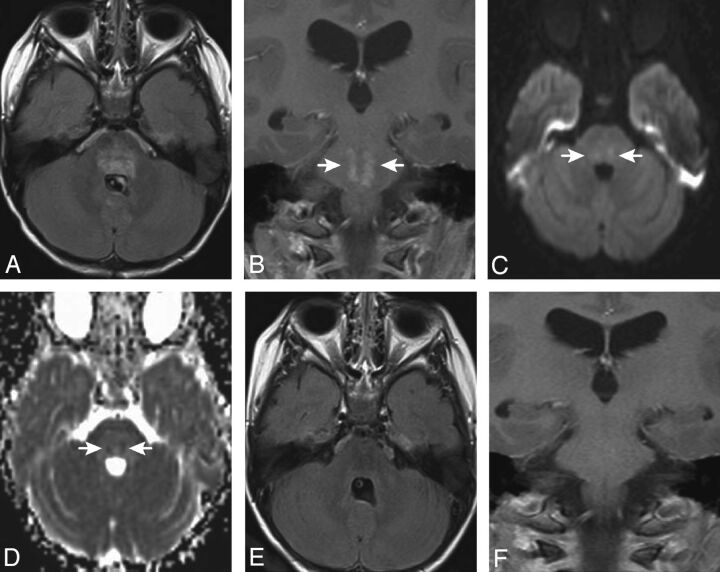
Fig 2.
Axial FLAIR (A) and postgadolinium coronal T1-weighted (B) images for patient 6, obtained 4.2 months after completion of PT, demonstrate patchy hyperintensity (FLAIR) and focal irregular enhancement (arrows in B) within the pons. Diffusion-weighted (C) and apparent diffusion coefficient map (D) images reveal small areas of restricted diffusion in the areas of abnormal signal and enhancement. This portion of the pons was in the high-dose volume of irradiation. The abnormalities improved on an MR imaging study obtained 1.5 months later and were no longer visible 4 months after they first appeared (E and F).

