Figure 7.
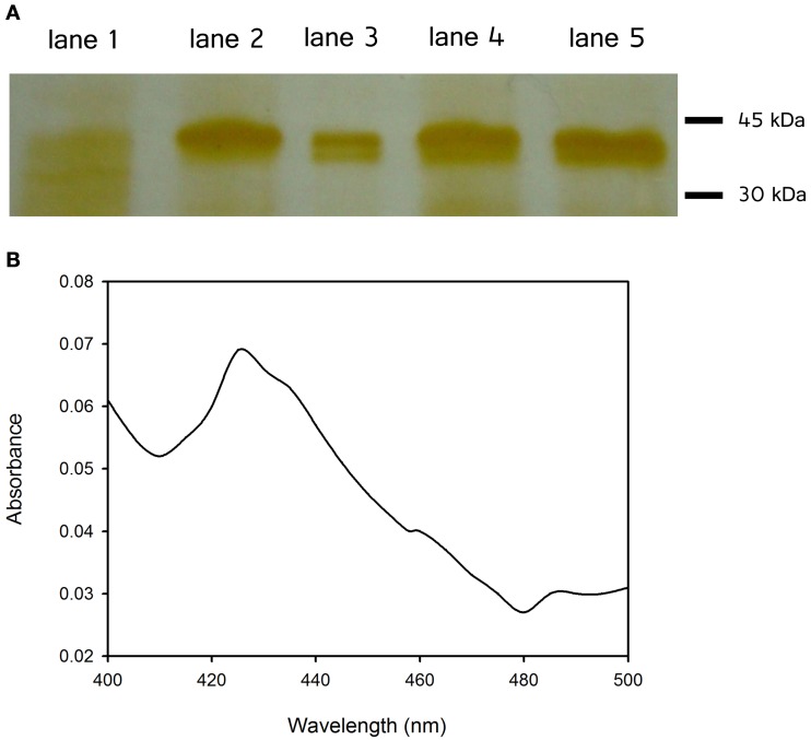
(A) SDS–PAGE of partial purified proteins using immobilized metal ion chromatography (Ni2+ IMAC): vector without insert (lane 1), CYP52X1 (lane 2), CYP5337A1 (lane 3), CYP617N1 (lane 4), and CYP53A26 (lane 5). Examined proteins were expressed in the yeast Saccharomyces cerevisiae WAT11. The cells were transformed with the lithium acetate method (Ito et al., 1983) for microsome isolation purposes. Proteins were solubilized with 10% w/v sodium cholate and loaded onto the column. (B) CO difference spectrum of CYP53A26: Sodium dithionite and CO were added to the purified protein, and absorbance between 400 and 500 nm was measured.

