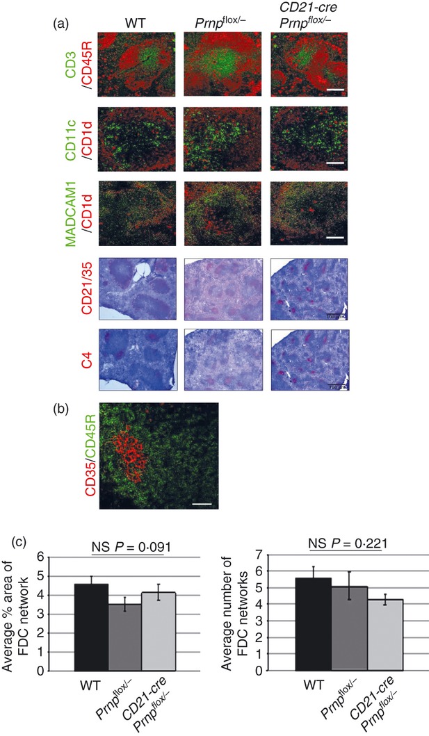
Figure 2.

Ablation of the cellular isomer of the prion protein (PrPC) specifically in follicular dendritic cells (FDC) has no effect on FDC status or lymphoid tissue microarchitecture. (a) Frozen spleens from mice with PrPC ablated specifically in FDC (CD21‐Cre Prnpflox/−), were immunolabelled for FDC (C4, CD21/35), B‐lymphocyte subsets (CD45R and CD1d), T lymphocytes (CD3), classical dendritic cells (CD11c) and marginal zone cells (MADCAM‐1 and CD1d). Comparison of sections from PrPC‐ablated animals with Prnpflox/flox or wild‐type (WT) controls showed no differences in the tissue microarchitecture or distribution of these cell subsets within the spleen. Scale bars on fluorescent images are 50 μm. Scale bars on light microscopy images are 500 μm and sections are counterstained with haematoxylin, blue. (b) Immunohistochemical analysis shows FDC (CD35+ cells, red), in contrast to B cells (CD45R, green), specifically express high levels of CD35 in the spleen. (c) Morphometric analysis of FDC confirmed that there were no significant differences in the size or number of FDC networks in each group (FDC networks/field‐of‐view when viewed with the × 10 objective; n = 48 images analysed per group). Statistical analysis carried out using one‐way analysis of variance. Data are representative of three independent experiments.
