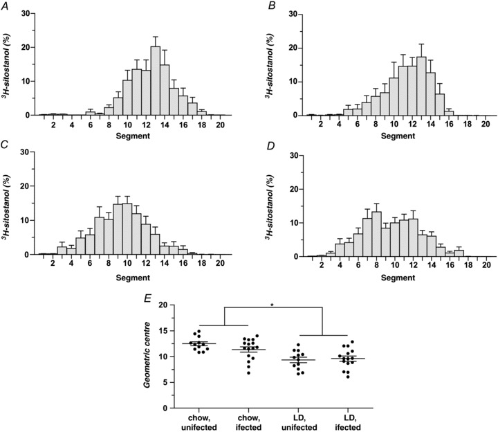
Figure 1. Influence of lithogenic diet (LD) and helicobacter infection on small intestinal transit times in male C57L mice.

A–D, small intestinal transit is plotted as per cent distribution of the non-absorbed marker 3H-sitostanol along the length of the small intestine 20 min after intra-duodenal injection (Time 0). Peak radioactivity on a chow diet occurs in segment 13 of uninfected mice (A) and H. hepaticus-infected mice (B). On the LD, peak radioactivity is found in segment 10 of uninfected mice (C) and in segment 8 of H. hepaticus-infected mice (D), indicating that LD feeding slows intestinal transit irrespective of infection status. E, small intestinal transit in the four groups is plotted as dimensionless geometric centres (Σ (fraction of 3H per segment × segment number)) of the radioactive marker for individual mice, with means ± SEM plotted for each group. The geometric centres of uninfected and infected mice on the chow diet (12.5 ± 0.4 and 11.4 ± 0.5, respectively) are significantly (P < 0.05) larger (i.e. intestinal transit is faster) than the geometric centres of the uninfected and infected groups fed the LD (9.4 ± 0.5 and 9.6 ± 0.5, respectively, as determined by two-way ANOVA). No statistically significant differences exist between the geometric centres of the two chow-fed or two LD-fed groups. *P < 0.05.
