
Figure 4. Effect of l-NNA and MRS2500 in the IJP induced by protocol 3 (5 Hz) of EFS.
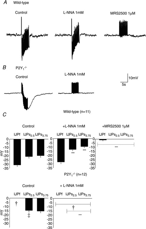
Representative recording of a wild type animal (A) and P2Y1−/− mouse (B). Notice the presence of a fast followed by a sustained component in the IJP recorded from colonic tissue of WT animals and the lack of the first fast IJP in P2Y1−/− mice. (Notice that positive deflections correspond to stimulus artefacts elicited at each frequency.) C, graph showing the amplitude of the response of the fast (IJPf) and slow (IJPs) component of the IJP (measured 2.5 and 3.75 s after the beginning of the stimulus) in WT animals (n= 11) and P2Y1−/− mice (n= 12). Comparison between the IJP amplitude from WT and P2Y1−/− mice was significant (two-way ANOVA: ‡P < 0.0001; †P < 0.0001 post hoc Bonferroni test). Comparison between the IJP amplitude after drug addition was also significant (in both cases: two-way ANOVA P < 0.0001) but MRS2500 (1 μm) was needed to abolish the IJP in WT animals. In contrast, no IJP was recorded after incubation with l-NNA in tissue from P2Y1−/− mice. ***P < 0.0001 post hoc Bonferroni test from previous drug addition.