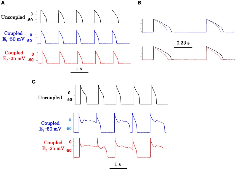
Figure 6.
Promotion of EADs by myofibroblast-myocyte coupling (M-F) during oxidative stress. Coupling a patch-clamped myocyte superfused with normal Tyrode's solution to a virtual fibroblast promotes no EADs (A). However, coupling lowers the AP plateau and shortens APD as shown in the superimposed traces at a faster time scale in panel (B). Panel (C) (top traces) shows lack of EAD induction with 0.1 mmol/L H2O2 in the uncoupled state. However, coupling the myocyte to a virtual fibroblast (Cf 6.3 pF, Gj 3.0 nS) caused EADs to reappear, which were more prominent when Ef was −25 mV (bottom traces) than −50 mV (middle traces). Cf is fibroblast capacitance in pF, Gj is M-F gap junctional conductance in nS, and Ef is the resting potential of the fibroblast in mV (from Nguyen et al., 2012).

