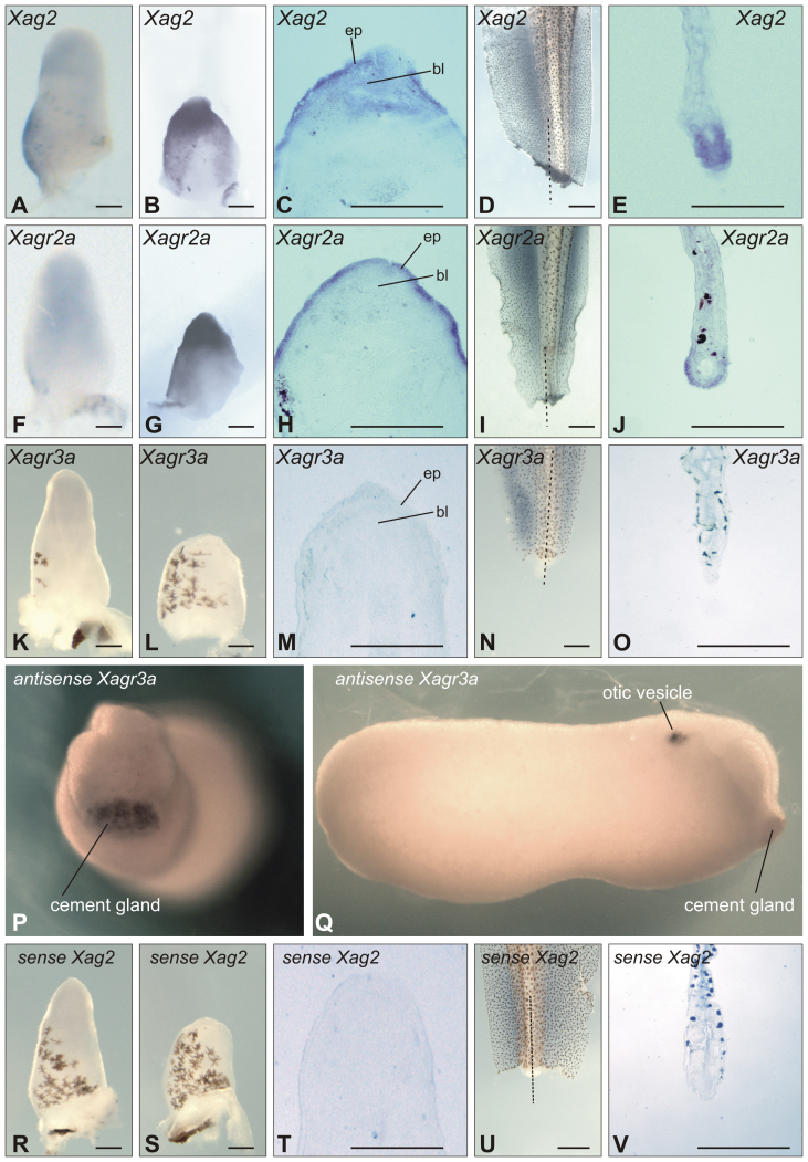
Figure 4. Analysis of Agrs expression by whole-mount in situ hybridisation.
(A) and (B), (F) and (G), (K) and (L). Intact hindlimb buds and hindlimb buds amputated at stage 52 and hybridised at 1 dpa with Xag2, Xagr2a and Xagr3a probes, respectively; distal to the top, ventral side to the right. (C), (H) and (M). Sagittal sections of amputated hindlimb buds shown on (B), (G) and L; ep – epidermis, bl – blastema. (D) and (E), (I) and (J), (N) and (O). Left side view and frontal sections (the level of section is indicated by dotted lines on (D), (I) and (N)) of tails amputated at stage 52 and hybridised at 1 dpa with Xag2 and Xagr2a probes; distal to the bottom, ventral side to the left. (P) and (Q). Stage 25 tailbud embryo hybridized with Xag3a antisense probe. Frontal and right side view respectively. Dorsal to the top. Anterior on B to the right. (R) and (S). Intact and amputated hindlimb buds operated at stage 52 and hybridized at 1 dpa with Xag2 sense (control) probe; distal to the top, ventral side to the right. (T). Sagittal sections of amputated hindlimb buds operated at stage 52 and hybridized at 1 dpa with Xag2 sense (control) probe. (U) and (V). Left side view and frontal sections (the level of section is indicated by dotted line on (U) of amputaited tail operated at stage 52 and hybridized at 1 dpa with Xag2 sense (control) probe; distal to the bottom, ventral side to the left. Bars – 250 microns.

