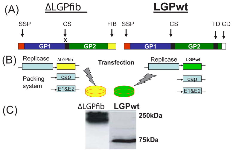
Figure 5. Generation of LASV GP VLPV.
(A) ΔLGPfib, C-truncated non-cleavable GP fused with T4 fibritin (FIB); LGPwt, “wild”-type GP: SSP, the stable signal peptide (aa 1–58); GP1 (59-259) and GP2 (260-491) subunits; CS, cleavage site for the SKI-1/S1P. The fusion peptide and transmembrane domain of the GP2 are shown in black boxes; CD, cytoplasmic domain. (B) GP genes were cloned into alphavirus vector and packed in VLPV (see text). (C) Western blot of cell extracts from Vero cells treated with VLPV (>2 VLPV/cell). 24 hours after treatment (exposure), cells lysates were prepared for SDS/PAGE. Samples were not boiled and run on 8% gel under non-reducing conditions. Proteins were blotted onto nitrocellulose membranes, treated with mAbs against LASV GP1 and virus-specific proteins were detected as previously described [45]. The GP1 ran off the gel under these gel electrophoresis conditions; 75 and 250kDa are proteins markers.

