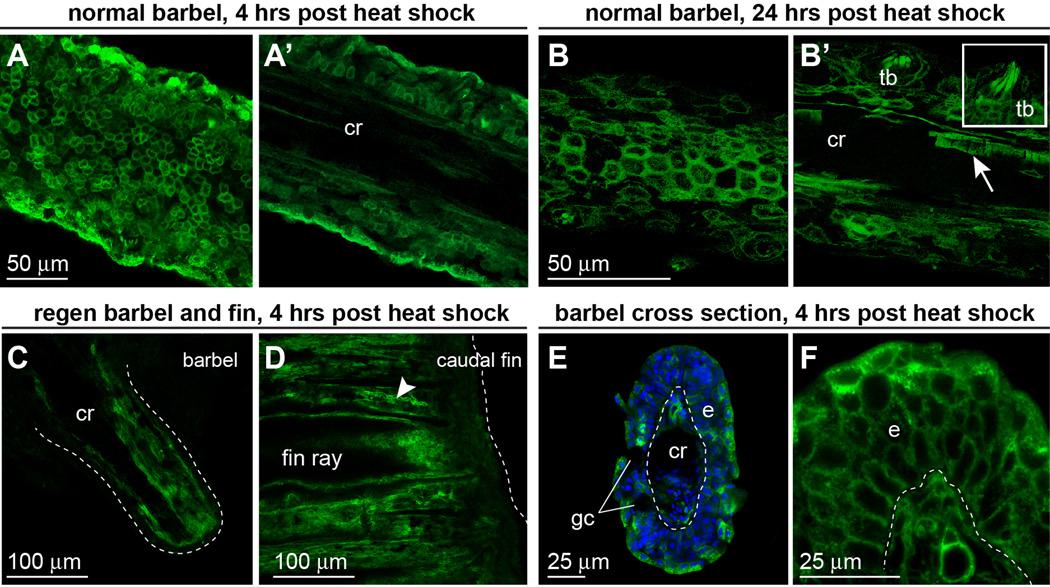
Figure 7. dn-fgfr1:EGFP is expressed throughout the maxillary barbel up to 24 hours after heat shock treatment.
A) Confocal image of an unfixed dn-fgfr1 maxillary barbel collected 4 hours after a one-hour heat shock. EGFP is prominent in epithelial cell membranes. A’) A deeper slice from the same z-stack shows EGFP in all cells surrounding the acellular central rod (cr). B). 24 hours after heat shock, membrane localization of EGFP is maintained in the barbel epithelium. B’) A deeper slice through the same z-stack shows persistent EGFP in basal epithelial cells, taste buds (inset), nerve fibers, and individual endothelial cells (arrow). C) Confocal slice through the center of an unfixed, amputated maxillary barbel 24 hours after heat shock; the distal end is to the lower right. EGFP is expressed throughout the full thickness of the stump, particularly in dermal tissues surrounding the central rod. Similar levels of EGFP expression were observed in amputated caudal fins at the same time point (D); the dotted line indicates the distal margin of each appendage. E) Fixed maxillary barbel tissue collected 4 hours after heat shock and mounted ‘on end’ in low-melt agarose. EGFP is visible in both epidermal (e) and deeper layers. Regions without EGFP include the epidermal goblet cells (gc) and the central rod (cr). F) ‘On end’ view of a second maxillary barbel showing membrane-localized EGFP in similar areas.

