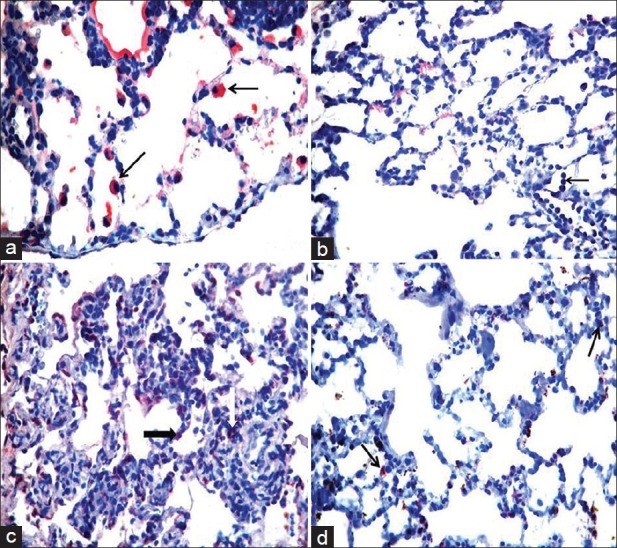
Figure 5.

Immunohistochemical examination (H and E, ×400). (a) Mainly positive staining for isoform of nitric oxide synthase (iNOS) in type II pneumocytes for control group (black arrows), (b) Normally marked myeloperoxidase (MPO) staining in neutrophils (black arrow) for control group, (c) Negative staining for iNOS in heliox group, (d) Slightly decreased MPO staining in heliox group
